Links | About | Simon Moyes | Contact
- Surgeons site
- Treatment
- Historical Development
- Advantages and Contraindication
- Anatomy
- Instrumentation
- Theatre Layout
- Diagnostic Ankle Arthroscopy
Quick Search
Ankle arthrodesis
An ankle arthrodesis, if successful, allows patients to return to work and some sports with a virtually normal gait.
Fusion rates have been reported from many series as in the order of 80% and infection occurring in 5-25% (103-107). Morgan in 1985 reported a 96% fusion rate with 90% good / excellent results; he maintained the contour of the talar dome, kept the ankle in neutral and used cross-screw internal fixation (108). Two years earlier, Schneider first described arthroscopic ankle arthrodesis (109). But it was Morgan (110) who published the first report in 1987. Myerson (111, 112) compared open and closed techniques of ankle arthrodesis with a reported quicker fusion time arthroscopically of 8.7 versus 14.5 weeks theoretically because of the lack of diruption pf the soft tissues and therefore a better blood supply to the fusing surfaces. The faster fusion rate was backed up by Ogilvie-Harris (113) who reported an 89% fusion rate arthroscopically with 88% fused by the third post operative month. Winson I G et al (114) reported a very large series of 118 arthroscopic fusions. The mean time to union was 12 weeks; non-union occurring in 7.6%.
The advantages of an arthroscopic arthrodesis are reduced morbidity, shorter hospital stay, faster fusion rate, better cosmesis and lower complication rates. Against these are a long learning curve for the surgeon and theatre staff, it can be a longer procedure and requires expensive arthroscopic equipment. Also it cannot correct large varus or rotational deformities.
The contra-indications for an arthroscopic arthrodesis are >15 degrees deformity, a previously failed arthrodesis, the presence of infection, RSD and a charcot joint.
Mann showed that the best fusion position is with the ankle in neutral, avoiding >10 degrees plantar-flexion and with the os-calcis in 5 degrees valgus (115). Also the ‘Mann’ position results in the best gait (116). You do, however, lose 70% of the total motion arc with an ankle fusion, and tarsal hypermobility is increased 85% (117).
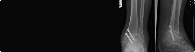
The arthroscopic technique is to have the standard arthroscopic set up with either invasive or non-invasive distraction. Remove all articular cartilage initially from the talar dome and plafond, then the gutters to expose bleeding underlying bone and finally the anterior osteophyte needs removal as this would otherwise resist talar reduction. Then the fusion is secured with parallel cannulated screws. Screw positioning is arthroscopically assisted and the length of the screws can be image intensifier assisted.
Patients then spend 3 weeks non weight bearing followed by 4-6 weeks partial weight bearing. The screws can be removed later if they are causing pain. A range of 3-12 months has been reported for standard open fusion to occur (118-120), this compares unfavourably with the arthroscopic technique. Mann (121) from a multi-centre trial recently demonstrated a 91% fusion fusion and 84% good / excellent results. This fusion rate leaps to 96% if known poor techniques are avoided, eg laser, external charnley type compression.
Click here for a video of ankle arthrodesis
Click here for information on rehabilitation
References
(103) Ahlberg A, Henricson A S, ‘Late results of ankle fusion’ Acta Orthop Scand 198; 52:103
(104) Boobyer G N, ‘The long term results of ankle arthrodesis’ Acta Orthop Scand 1981; 52:107
(105) Johnson F W, Boseker E H, ‘Arthrodesis of the ankle’ Arch Surg 1968; 97:766
(106) Morrey B F, Wiedeman G P, ‘Complications and long term results of ankle arthrodesis following trauma’ JBJS 1980; 62A777
(107) Said E, Hunka L, Siller T M, ‘Where ankle fusion stands today’ JBJS 1978; 60B:211
(108) Morgan C D, Henke J A, Bailey R W, Kaufer H, ‘Long term results of tibio-talar arthrodesis’ JBJS 1985; 67A:546
(109) Schneider D, ‘Arthroscopic ankle fusion’ Arth Video 1983; 3
(110) Morgan D C, ‘Arthroscopic tibiotalar arthrodesis . Jefferson Orthop J 198; 16:50
(111) Myerson M S, Allon S M, ‘Arthroscopic ankle arthrodesis . Contemp Orthop 1989; 19:21
(112) Myerson M S, Quill G, ‘Ankle arthrodesis - a comparison of an arthroscopic and an open method of treatment’ Clin Orth 1991; l 268:84
(113) Ogilvie-Harris D J, Lieberman I, Fitsialis D, ‘Athroscopically assisted arthrodesis for osteoarthritic ankles’ JBJS 1993; 75A 1167
(114) Winson I G et al, ‘Arthroscopic arthrodesis’ JBJS 2005; 87:3; 343-7
(115) Mann R A, Coughlin M, ‘Surgery of the foot and ankle’ St.Louis: C V Mosby, 1991; 676
(116) Mazur J M, Schwartz E, Simon S R, ‘Ankle arthrodesis: long term follow-up with gait analysis’ JBJS 1979; 61A:964
(117) Morgan C D, Henke J A, Bailey R W, Kaufer H, ‘Long term results of tibio-talar arthrodesis’ JBJS 1985; 67A:546
(118) Campbell C J, Rinehart W T, Kalenak A, ‘Arthrodesis of the ankle: deep autogenous graft with maximum cancellous bone apposition’ JBJS 1974; 56A:63
(119) Scranton P E Jr, ‘Use of internal compression in arthrodesis of the ankle’ JBJS 1985; 67A:550.
(120) Holt E W, Hanson S T, Mayo K A, Sangeorzan B J, ‘Ankle arthrodesis using internal screw fixation’ Clin Orth 1991; 268:21
(121) Mann J A, Glick J M, Morgan C T et al, ‘Arthroscopic ankle arthrodesis: experience with 75 cases’ AAOS Feb 1995